
PRODUCT
Home / Product




[리퍼비시] BioTek Lionheart LX Automated Microscope
Lionheart LX 자동화 현미경은 경제적으로 설계되었으며, 학습 및 사용이 복잡한 고가의 맞춤 제작된 자동화 현미경에 대한 대안을 제공합니다. 콤팩트하며 접안 렌즈가 없는 하드웨어 설계는 사용이 더욱 편리하며 작업자의 인체공학적 고통을 줄여줍니다.
Lionheart LX는 Gen5 소프트웨어와 함께 이미지 캡쳐, 처리 및 분석을 완전히 자동화하는 Augmented Microsopy입니다. 시료를 넣고 실행하도록 작동이 쉽고, 즉시 게재 가능한 이미지와 정량분석 데이터를 수집합니다. Lionheart LX는 연구 성과를 높이고 시간과 비용을 절감할 수 있습니다.
기술사양
- 1.25배~60배 공기
- 60배~100배 오일 이멀전
- 4배~40배 위상차
- 조이스틱 스테이지 컨트롤
- 레이저 자동 초점
- 광시야 형광
- 명시야
- 고대비 명시야
- 컬러 명시야
- 위상차
- 단일 컬러
- 멀티 컬러
- 몽타주
- 타임랩스
- Z-스태킹
- 6~1536-well plate
- 슬라이드
- 페트리/세포 배양 접시
- 세포 배양 플라스크(T75 및 T25)
- 혈구계(Hemocytometer)
- 챔버 슬라이드
- 도립 현미경
• Lionheart LX는 설치 공간이 작고 대부분의 표준 실험실 벤치에 쉽게 설치할 수 있습니다. 과도한 장비나 암실이 불필요
• Lionheart는 고품질 Olympus 대물렌즈와 Semrock 필터를 사용하여 뛰어난 이미지 품질과 성능을 제공합니다
• 넓은 시야각(WFOV) 카메라는 연구자들이 필요한 답을 빠르게 얻을 수 있도록 마이크로플레이트와 슬라이드에서 빠르고 자동화된 이미징 기능을 제공합니다
• Lionheart LX는 Gen5 이미징 소프트웨어와 함께 사용될 경우, 독보적인 완전 자동화 캡처, 처리, 분석, 출판 워크플로를 제공합니다
• 레이저 기반 및 소프트웨어 기반 자동 초점은 자동화된 현미경에서 정확한 초점의 선명한 이미지를 캡처하는 데 필수적입니다
• Lionheart LX 스테이지의 오픈형 설계는 연구자들로 하여금 마이크로유액 장비를 사용할 수 있도록 합니다
| 배율 범위 |
|
| 이미저/리더 옵션 |
|
| 이미징 모드 |
|
| 이미징 방법 |
|
| 지원되는 랩웨어 |
|
| 현미경 유형 |
|
특징
Small benchtop footprint
The Lionheart FX has a small footprint and is easily installed on a standard lab bench with no need for a dedicated darkroom.
Wide field of view
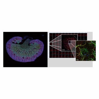
The wide field of view (WFOV) camera provides fast, automated imaging in microplates and slides. Large tissue sections (left) and microplates (right) can be imaged from low to high magnifications.
Autofocus customized for your sample types and labware
A. Image-based autofocus is available on all Agilent BioTek imaging systems. It focuses on the plane of highest contrast in the sample, including “shifting” biology within the well.
B. The BioTek proprietary Laser autofocus method uses the same focal offset from a laser identified interface unique to each well or area to be imaged. It is typically faster, works with dim fluorophores, and helps prevent phototoxicity and photobleaching. Laser autofocus also offers better reproducibility and higher accuracy during long term kinetic imaging.
Augmented microscopy
When paired with Gen5 imaging software, the Lionheart LX provides a unique fully automated capture, process, analyze and publish workflow.
Open-stage design
The open design of the Lionheart LX stage allows researchers to work with microfluidic devices.
응용분야
Proprietary Laser Autofocus
Laser autofocus and image-based (or software-based) autofocus are two methods of autofocus widely used in microscopy.
Cell Counting
Accurate cell counting is a critical step across a spectrum of cell biology applications. Direct cell counting uses methods that identify and enumerate individual cells, as opposed to indirect methods that estimate population sizes using biochemical methods.
Object Tracking
With the incorporation of the object tracking module, available within Agilent BioTek Gen5 software for imaging and microscopy, this can be easily accomplished in a fully automated fashion.
Automatic Region of Interest Identification
This process enables walk-away slide scanning for four or more slides, if combined with Agilent automation offerings, and can be used with the Agilent BioTek Lionheart automated microscopes and Agilent BioTek Cytation imaging multimode readers.
Spot Counting
The spot counting module available in Agilent BioTek Gen5 software enables the number of spots counted, and other information, to be tied to the original primary mask data, allowing these values to be known for each object initially identified in the image.
Augmented Microscopy
Agilent BioTek instrumentation and software create the unique augmented microscopy experience with the integration and automation of all steps from image capture to publication-ready data.